Mohs surgery for melanoma in situ now offered at Cary Skin Center
Cary Skin Center is now using the Mohs technique for melanoma in situ
An estimated 76,000 people in the United States were diagnosed with melanoma in 2016. Melanoma is potentially one of the most dangerous of all skin cancers, and its incidence is rising steadily. Even when melanoma is in situ — an early stage during which the cancer has not yet grown below the external layer of skin — removing it is often a multistage process that takes a few days as patients and doctors wait for tissue analysis. Without the finesse of a layer-by-layer surgery called Mohs, which is used universally for other forms of skin cancer, removal of early-stage melanoma can produce large wounds and disfiguring scars.
This March Cary Skin Center will begin using the Mohs technique for melanoma in situ. This outpatient surgery procedure has been reported to be less expensive than the traditional surgical approach, creates a smaller wound and reduces the cancer’s rate of recurrence. A melanoma in situ can be removed and reconstructive surgery completed all in one day.
Using Mohs to treat melanoma in situ became possible when researchers figured out how to use special stains with tissue samples of the cancer. Unlike more common forms of skin cancer, such as basal cell or squamous cell carcinoma, melanoma’s cells are difficult to visualize with the standard dyes used to prepare slides for analysis. About 15 years ago, researchers finally developed an immunohistochemistry stain for melanoma, tuned to the cancer’s particular biomarkers. That discovery made melanoma visible, but initially it was only used for formalin slides, which take about 12 hours to prepare, not the quicker, frozen-section slides used currently in Mohs surgery.
In the past, Mohs was rarely chosen for melanoma surgery for fear that some microscopic melanoma cells might be missed and end up metastasizing. In recent years, however, efforts to improve and refine the Mohs surgeon’s ability to identify melanoma cells have resulted in the development of special stains that highlight these cells. These special stains are known as immunocytochemistry or immunohistochemistry (IHC) stains and use substances that preferentially stick to melanocytes (the cells that become melanoma) making them much easier to see with the microscope.
Doctors wanted to do with melanoma what they could do with other skin cancers: remove a tissue sample that could be stained, frozen and slide-ready in less than an hour. Clinicians were hesitant to use the new stain in the quick-processing preparation until studies showed it was as accurate as the previous formalin-slide-based, traditional analysis.
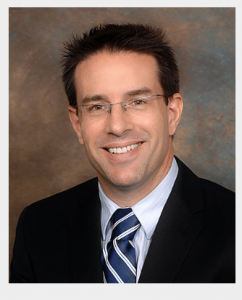
That evidence was published in the Journal of the American Academy of Dermatology. “The published study added to the growing body of evidence that shows the incredible usefulness of this immunohistochemical marker in frozen-section analysis in Mohs for in situ melanoma,” said surgeon Adam Ingraffea MD, Mohs surgeon at Cary Skin Center. “We are incredibly pleased about those results and about Cary Skin Center’s ability to now offer this new technique to our patients.”
The Mohs procedure is based on a technique developed by Wisconsin surgeon Frederic Mohs in the 1930s. Mohs surgery begins with the removal of the most visible center of the cancer. Then, the surgeon will remove tissue around the center, in layers millimeters thick. That tissue is sliced into micron-thin pieces, stained, frozen and examined within minutes. The incremental removal of tissue means that no more will be removed than is necessary. When the cancer is near the eyelid, for instance, the precision of Mohs keeps wounds and scars, or reconstructive surgery, to a minimum.
Dr. Adam Ingraffea and fellow dermatologic surgeon Elias Emile Ayli, DO, expect to use the procedure to treat the increasing number of people diagnosed with melanoma. The new technique is well-suited for patients whose melanoma is in areas where sparing tissue is most critical —such as the head and neck. With speedy analysis of cancerous tissue now available, patients can have a tumor removed and any needed wound repair completed in one day.
About Cary Skin Center:
In 1998, Dr. Robert E. Clark established the Cary Skin Center, a state-of-the-art outpatient surgical center specializing in Mohs Micrographic Surgery for the removal of skin cancer. Dr. Timothy C. Flynn joined the Cary Skin Center in 2001 and together they have successfully treated countless patients in the Triangle and surrounding areas.
Because Mohs surgery is a highly complex and sophisticated surgical method, it requires extensive training. Dr. Clark, Dr. Flynn, Dr. Ayli and Dr. Ingraffea completed 1-2 year intensive training programs, including complex surgical cases and advanced reconstruction to become Fellows of the American College of Mohs Surgery. In addition, they offer over 60 years of combined Mohs surgery experience.
Parker Eales
Cary Skin Center
919-303-4053
email us here
WHO TO TRUST TO DO YOUR SKIN CANCER TREATMENT
EIN Presswire does not exercise editorial control over third-party content provided, uploaded, published, or distributed by users of EIN Presswire. We are a distributor, not a publisher, of 3rd party content. Such content may contain the views, opinions, statements, offers, and other material of the respective users, suppliers, participants, or authors.


